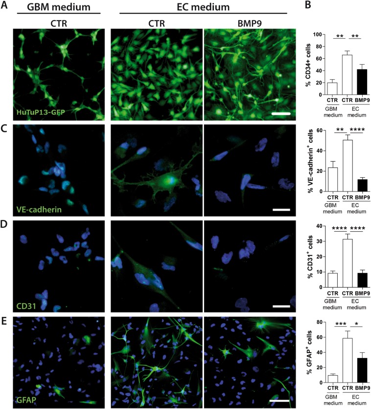
Fig. 4.
Endothelial commitment is impaired by BMP9. Representative images showing cell morphology of GFP-transduced cells (HuTuP13) affected by EC medium and BMP9 treatment at 30 ng/ml every other day for 10 days (original magnification 10×, scale bar = 100 μm) (a). Flow cytometry analysis of CD34+ after 10 days of treatment with BMP9 at 30 ng/ml every other day (HuTuP13/83/108/175) (b). Representative images (HuTuP174) of immunofluorescence staining for VE-cadherin (green, c), CD31 (green, d), GFAP (green, e) and relative quantifications (right panels), after 10 days of treatment at 30 ng/ml every other day (original magnification 20×, scale bar = 50 μm). Data are presented as mean ± S.E.M. of N ≥ 3 independent experiments. *p < 0.05; **p < 0.01; ***p < 0.001; ****p < 0.001 by paired t-test or One-way ANOVA

