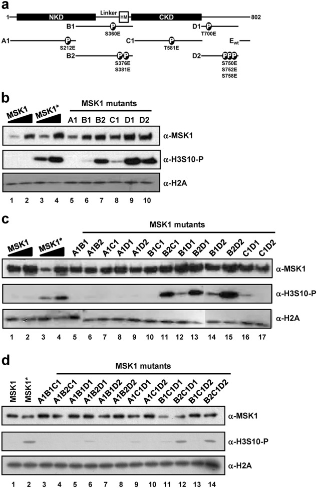
Fig. 4. Characterization of histone phosphorylation activities of MSK1 phosphomimetic mutants.

a Schematic diagram of MSK1 with the N-terminal kinase domain (NKD), hydrophobic motif (HM) in the linker region and C-terminal kinase domain (CKD). Phosphomimetic mutations are indicated in the A1 (S212E), B1 (S360E), B2 (S367E and S381E), C1 (T581E), and D1 (T700E) and D2 (S750E, S752E, and S758E) fragments. b–d Reactions containing core histone octamers and purified MSK1 with a single (b), double (c), and triple (d) number of mutant fragments were subjected to in vitro kinase assays. The kinase activities of purified MSK1 were measured by immunoblot with anti-phosphorylated H3S10 antibody. MSK1 and MSK1* wild-type MSK1 were prepared in the absence and presence of MKK6ca co-expression, respectively. The data are representative of at least three independent experiments
