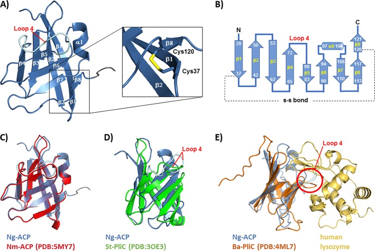
FIG 2.
Structure of rNg-ACP. (A) Cartoon representation of the three-dimensional structure of rNg-ACP, showing the antiparallel arrangement of eight β-strands. The position of the stabilizing disulfide bond is shown in stick representation in the zoomed view. (B) Topology diagram showing residue ranges in each β-strand and the -S-S- bond between Cys37 and Cys120 residues. (C) Superposition of Ng-ACP (PDB code 6GQ4, blue) with Nm-ACP (PDB code 5MY7, red). (D) Superposition of Ng-ACP (PDB code 6GQ4, blue) with S. enterica Typhimurium PliC (St-PliC; PDB code 3OE3, green). (E) Superposition of Ng-ACP (PDB code 6GQ4, blue) with the Brucella abortus PliC (Ba-PliC; PDB code 4ML7, orange)-lysozyme complex (gold); the position of loop 4 is indicated by the red circle. Visualization of structures was done with PyMOL.

