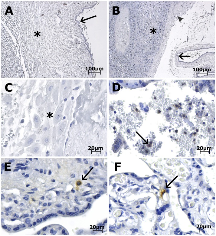
Figure 3.
Photomicrography of placenta, cord and membrane samples from women diagnosed as positive for ZIKV infection immunostained with anti-ZIKV MAb and stained with Harris's hematoxylin. The scale bars are 100 μm (A,B) and 20 μm (C–F). (A) Case LRV/16 515—Umbilical cord slides from a third trimester placenta negative for anti-ZIKV MAb. Fibroblasts in Wharton's jelly (*) and amniotic epithelium (arrow) are negative for the antibody used. (B) Case LRV/16 515—Chorioamniotic membrane from a third trimester placenta negative for anti-ZIKV MAb. Notice that the capsular decidua (*), the smooth chorion above the capsular decidua (arrowhead) and the amniotic epithelium (arrow) are negative for the antibody used. (C) Case LRV/16 515—Decidua basalis (*) from a third trimester placenta negative for anti-ZIKV MAb. (D) Case LRV/15 572—Intervillous space from a first trimester placenta with inflammatory cell infiltrates positive for anti-ZIKV MAb (arrow). (E) Case LRV/16 927 and (F) Case LRV/16 848: Chorion frondosum from third trimester placentas showing Hofbauer cells positive for anti-ZIKV MAb (arrows) inside the chorionic villi. Notice that the cytotrophoblast and syncytiotrophoblast cells, as well as fibroblastic cells inside Wharton's jelly, are negative for the antibody used.

