
Fig. 1.
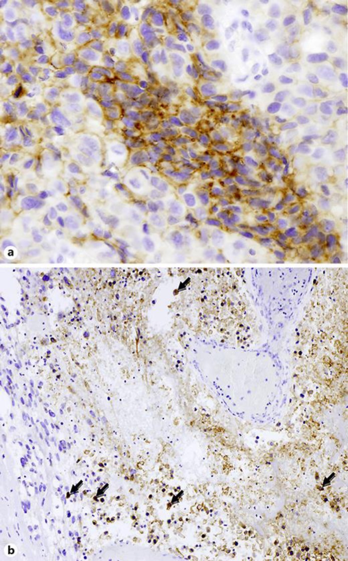
Immunohistochemistry for PDL-1 (clones SP263 by Ventana Medical System and E1L3N by Cell Signaling) showing a strong expression in both tumoral cells (a, tumor proportion score ≥50%; original magnification ×200) and inflammatory cells inside an area of tumor necrosis (b, macrophages and lymphocytes with arrows; original magnification ×100).