Figure 1:
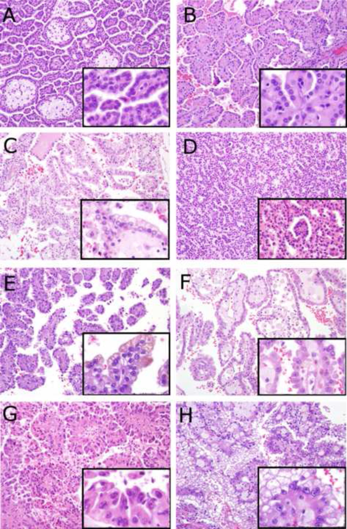
Microphotographs - larger photographs 200× magnification, smaller inset photographs 400× magnification of the same tumor. A – Classic type 1 papillary renal cell carcinoma with low grade (Fuhrman grade 2) nuclei, amphophilic cytoplasm, papillary architecture, and foamy macrophages within papillae. B – Type 1 papillary renal cell carcinoma with low grade (Fuhrman grade 2) nuclei and abundant eosinophilic cytoplasm. C – Type 1 papillary renal cell carcinoma with Fuhrman grade 2 nuclei and optically clear cytoplasm. D – Type 1 papillary renal cell carcinoma with Fuhrman grade 2 nuclei and glomerulations. E – Type 1 papillary renal cell carcinoma with higher grade (Fuhrman grade 3) nuclei. F – Type 1 papillary renal cell carcinoma with higher grade (Fuhrman grade 3) nuclei and abundant eosinophilic cytoplasm. G – Classic type 2 papillary renal cell carcinoma with higher grade (Fuhrman grade 3) nuclei, true pseudostratification, and abundant eosinophilic cytoplasm. H – Representative image from a Type 2 papillary renal cell carcinoma which demonstrated papillary areas with cells with eosinophilic cytoplasm (not shown in H) intermixed with other areas where the cytoplasm was more optically clear (which are demonstrated here).

