
FIGURE 8:
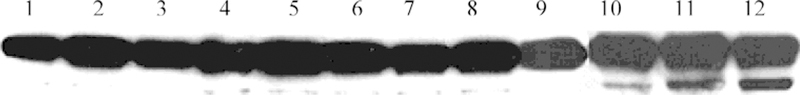
Different degrees of PARP cleavage in fully transformed (3Y1v-Src), partially transformed (3Y1c-Src), and normal (3Y1) rat fibroblasts as indications of apoptosis. 3Y1, 3Y1c-Src, and 3Y1v-Src cells were treated with 8 µM P-Glu4 for 24 h. Cells were irradiated at 0.84 mW cm−2 for 3.5 min (1.76 kJ m−2), and 9 h later the cells were collected and lysed. The supernatant of the lysate was applied to Western blot to detect PARP cleavage. Lane 1: 3Y1 cells with no photodynamic treatment; lane 2: 3Y1c-Src cells with no photodynamic treatment; lane 3: 3Y1v-Src cells with no photodynamic treatment; lane 4: 3Y1 cells with irradiation but no P-Glu4; lane 5: 3Y1c-Src cells with irradiation but no P-Glu4; lane 6: 3Y1v-Src cells with irradiation but no P-Glu4; lane 7: 3Y1 cells with P-Glu4 but no irradiation; lane 8: 3Y1c-Src cells with P-Glu4 but no irradiation; lane 9: 3Y1v-Src cells with P-Glu4 but no irradiation; lane 10: 3Y1 cells with irradiation and P-Glu4; lane 11: 3Y1c-Src cells with irradiation and P-Glu4; and lane 12: 3Y1v-Src cells with irradiation and P-Glu4.