Figure 2.
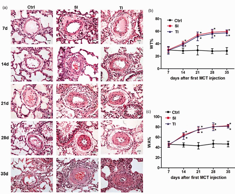
Monocrotaline (MCT)-induced structural changes and pulmonary vascular remodeling in lungs from SD rats. (a) Representative histological pictures correspond to SD rats that received normal saline (ctrl), single injection of 40 mg/kg MCT (SI group), and twice injections of 20 mg/kg MCT (TI group) on days 7, 14, 21, 28, and 35. Structure of pulmonary arteries with HE staining showed an intact pulmonary arterial wall and structure, without interstitial inflammatory cells in the control group. In contrast, inflammatory cells were observed in the interstitium on day 7 in both the SI and TI groups. These changes were aggravated further, along with impaired alveoli, and hyperplastic lung interstitium on days 14 through 35. (b, c) No changes in the percentage of total wall thickness to external diameters of pulmonary arterioles diameter (WT%) and the percentage of wall area to the total area of vessels (WA%) were observed on day 7 for all three groups. However, WA% and WT% of the pulmonary arteries were significantly increased in both the SI and TI groups vs. control on days 14, 21, and 28; whereas, no significant difference between the two injection groups was observed. On day 35, WA% and WT% in all groups remained similar to day 28. Scale bar = 25 μm. *P < 0.05 vs. ctrl of the corresponding day; #P < 0.05 vs. SI group.

