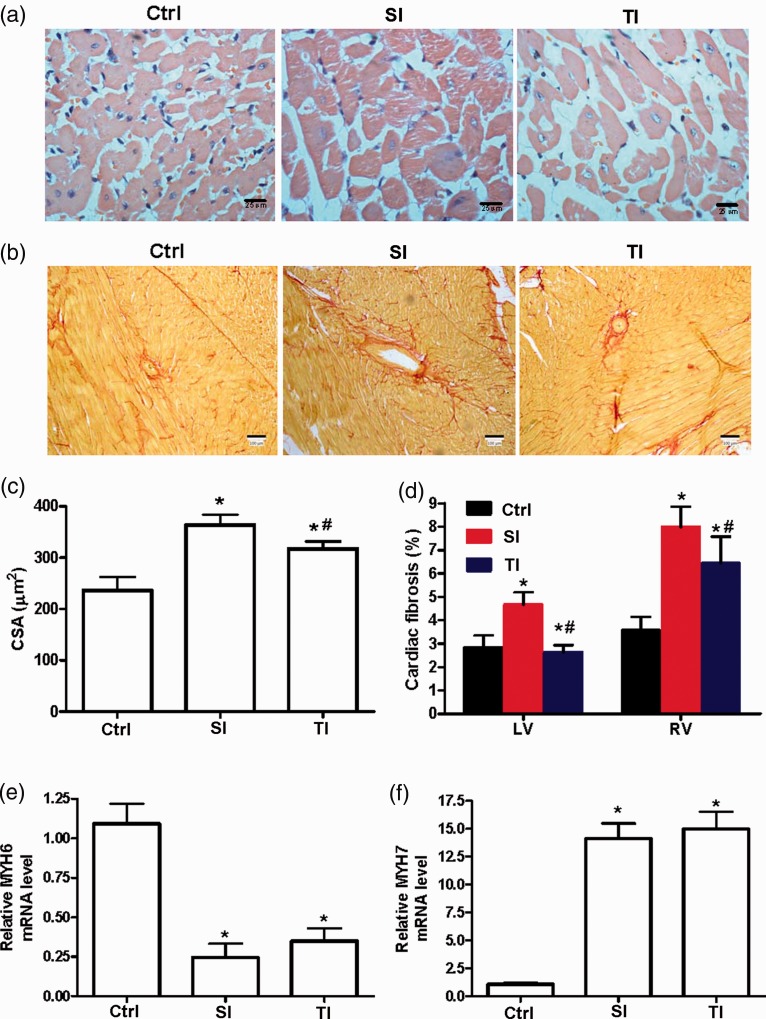
Figure 4.
Cardiac hypertrophy and fibrosis of rats. (a) Representative images of right ventricle (RV) sections from male rats stained with HE in ctrl, SI, and TI groups. (b) Representative images of RV fibrosis stained with picrosirius red staining. (c, d) Quantification of cardiomyocyte cross-sectional area (CSA, c), left ventricle and RV fibrosis (d) in different groups. (e, f) RV hypertrophy markers alpha myosin heavy chain (MYH6) and beta myosin heavy chain (MYH7) were measured by RT-qPCR. SI group: intraperitoneally injected with a single dose of 40 mg/kg monocrotaline (MCT); TI group: twice injections of 20 mg/kg MCT with an interval of seven days; ctrl: injected with normal saline. Scale bar = 100 μm. *P < 0.05 vs. ctrl of the corresponding day; #P < 0.05 vs. SI group.

