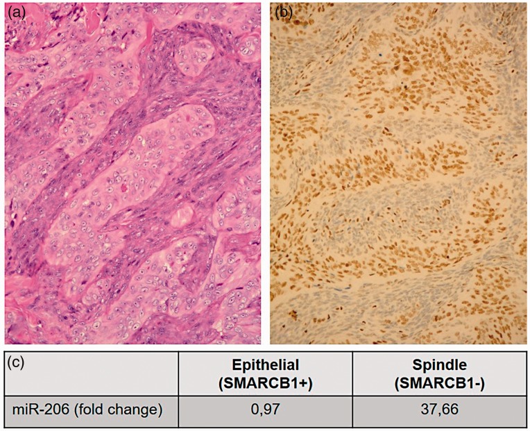
Figure 1.
Classical hematoxylin and eosin (H&E) staining (a) and SMARCB1 immunhistochemistry (b) of a biphasic synovial sarcoma. Epithelial components show positive reaction with SMARCB1 immunostaining, while the spindle cell components are negatively stained (b). miR-206 is highly overexpressed (37,66-fold) in the spindle cell components compared to the epithelial components, measured with q-RT-PCR. Relative miR-206 level was normalized to endogenous RNU6B. As calibrator, normal liver tissue was used (c). (A color version of this figure is available in the online journal.)

