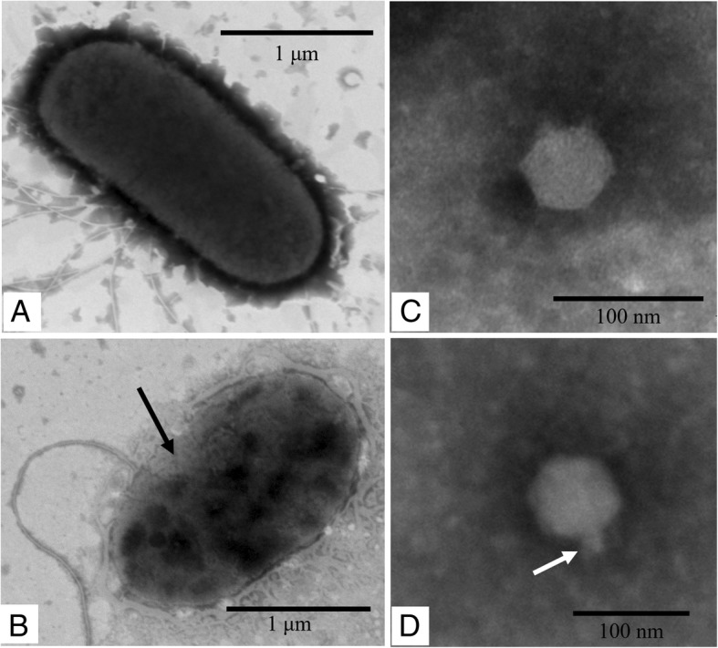
Fig. 1.

Electron micrograph of V. alginolyticus and phage Vp670. a A normal V. alginolyticus cell. b An infected V. alginolyticus cell on the verge of lysis. Numerous phage particles (shown by a black arrow) have been assembled within the cell. c The morphology of phage Vp670. d The short tail of phage Vp670 is shown by the black arrow
