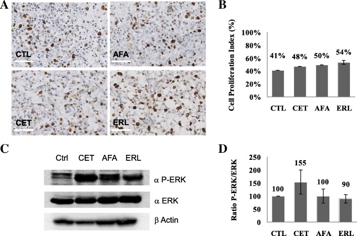
Fig. 4.
Inhibition of cell proliferation and the EGFR pathway in tumour explants. a) Microscope images (× 40) of Ki67 immunostaining on tumour slices from the patient’s glioblastoma for each condition: control (CTL), cetuximab (CET), afatinib (AFA), and erlotinib (ERL). b) Visual quantification of the proportion of Ki67-positive cells. There were no statistically significant differences between CTL on one hand and CET (p = 0.45), AFA (p = 0.6) and ERL (p = 0.37). c) Immunoblot of tumour slices treated for 48 h under four conditions: CTL, CET, AFA and ERL. To investigate the drugs’ efficacy, antibodies against phospho-ERK (P-ERK) and ERK (ERK) were applied, and actin was used as a loading control. d) Quantification of the P-ERK/ERK ratio, as a percentage of the control P-ERK/ERK ratio. There were no statistically significant differences between the negative control on one hand and cetuximab (p = 0.36), afatinib (p = 0.99) and erlotinib (p = 0.59) on the other. Values are expressed as the mean (n = 4) and the standard error of the mean (represented by error bars). The scale bar corresponds to 25 μm

