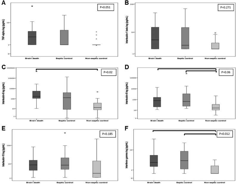
Fig 1.
Plasma cytokine values determined by magnetic bead assay in brain-dead patients and controls. A. Tumor necrosis factor (pg/mL). B. Interleukin-1β (pg/mL). C. Interleukin-6 (pg/mL). D. Interleukin-8 (pg/mL). E. Interleukin-10 (pg/mL). F. Interferon-γ (pg/mL). Kruskal–Wallis with pairwise comparison. Statistically significant differences as indicated by the bars (IL-6: P = 0.01 for BD vs. non-septic controls; IL-8: P = 0.029 for BD vs. non-septic controls and P = 0.01 for septic vs. non-septic controls; IFN-γ: P = 0.028 for BD vs. non-septic controls and P = 0.031 for septic vs. non-septic controls). Graphs are plotted on a logarithmic scale, representing median and interquartile range. Dots and asterisks represent outliers.

