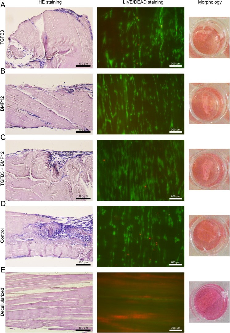
Fig 3.
Microscopic and macroscopic appearance of MSC-seeded tendon scaffolds and an unseeded control. Representative images of H&E-stained paraffin sections (left) of MSC-seeded scaffolds preloaded with TGFβ3 (A), BMP12 (B), TGFβ3, and BMP12 (C) or without any growth factor (D) (20x magnification; calibration marks correspond to 100 µm). Corresponding images of the LIVE/DEAD® staining (middle) and the macroscopic appearance of the scaffolds (right) are given. In fluorescence microscopic images of the LIVE/DEAD® staining presented in the middle panel, vital cells are indicated by green fluorescence (display of intracellular esterase activity), cells with defect cellular membranes show a red fluorescence signal of their nucleus (10x magnification; calibration marks correspond to 200 µm). Adequate images of unseeded control scaffold are added at the bottom to reflect the most likely cellular origin of morphological changes in MSC seeded scaffolds. All images were taken on day 5 from samples, which received MSC of one single donor.

