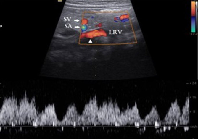
Figure 2.

Duplex Doppler sonography in longitudinal axis shows the shunt vessel (arrowhead) with pulsatile flow owing to the transmitted cardiac periodicity. Flow direction is towards the left renal vein. LRV, left renal vein,; SA, splenic artery; SV, splenic vein.
