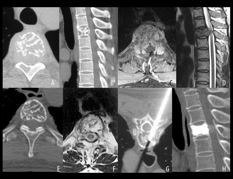
Figure 3.
A case of a 43-year-old man with mild myelopathy and pain for four months (Frankel grade of D) showing vertebral computed tomography (CT) and magnetic resonance imaging (MRI) and histopathology following radiotherapy for aggressive vertebral hemangioma (VH). A 43-year-old man experienced mild myelopathy and pain for four months (Frankel grade D). After percutaneous biopsy, he underwent radiotherapy with 40 Gy. Complete neurological recovery was achieved one year after radiotherapy with minor re-ossification, requiring vertebroplasty 60 months after radiotherapy because of a suspicion of a possible pathological fracture. (A) Axial computed tomography (CT) scan of an osteolytic lesion in the vertebral body and right lamina of T4. (B) Sagittal CT scan of an osteolytic lesion in the vertebral body and right lamina of T4. (C) Axial magnetic resonance imaging (MRI) showing epidural compression with vertebral canal stenosis (33.4%). (D) Sagittal MRI shows epidural compression with vertebral canal stenosis (33.4%). (E) Slight re-ossification on disappearance of epidural compression 60 months after radiotherapy. (F) Slight re-ossification upon disappearance of epidural compression 60 months after radiotherapy. (G) Negative pathology results following percutaneous biopsy and vertebroplasty. (H) Axial CT scan at the 108 months of follow-up showing no recurrence of the hemangioma.

