Figure 1.
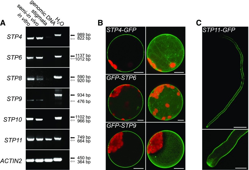
Analyses of Pollen Tube-Specific Expression of Different STPs and of the Subcellular Localization of the Encoded Proteins.
(A) RT-PCR-based comparison of STP4, STP6, STP8, STP9, STP10, and STP11 expression in in vitro-germinated pollen tubes, in pollen tubes grown through a stigma (semi-in vivo), and in virgin stigmata with gene-specific primers (Supplemental Table 3). Arrows indicate the predicted sizes of PCR products derived from genomic DNA (black) and reverse-transcribed mRNA (white). The presence of RNA in each sample was confirmed with ACTIN2 specific primers (Supplemental Table 3).
(B) Single optical sections (left) and maximum projections (right) of mesophyll protoplasts expressing GFP fusion constructs of STPs under the control of the 35S promoter. GFP is given in green and chlorophyll autofluorescence in red.
(C) Confocal optical section of a tobacco pollen tube transiently expressing STP11-GFP under the control of LAT52pro after particle bombardment. The bottom image shows the pollen tube tip at higher magnification. Bars = 10 µm in (B) and (C) (bottom) and 50 µm in (C) (top).

