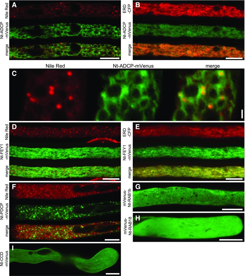
Figure 3.
Proteins Not Localizing to LDs.
The proteins were transiently expressed in tobacco pollen tubes, as N-terminal ([A] to [F] and [I]) or C-terminal ([G] and [H]) fusions to the fluorescent protein mVenus. The tubes were cultivated for 5 to 8 h, fixed with formaldehyde, and stained with Nile Red ([A], [C], [D], and [F]), only fixed ([B] and [E]), or not treated ([H] and [I]). Then, they were monitored by confocal microscopy. (C) is a magnified section of (A), showing partial association of mVenus and Nile Red fluorescence. Images are representative of 10 ([A], [C] to [E], and [H]), 14 ([B] and [I]), 7 ([C] and [F]), and 13 (G) pollen tubes. Bars = 10 µm in (A), (B), and (D) to (I) and 1 µm (C).

