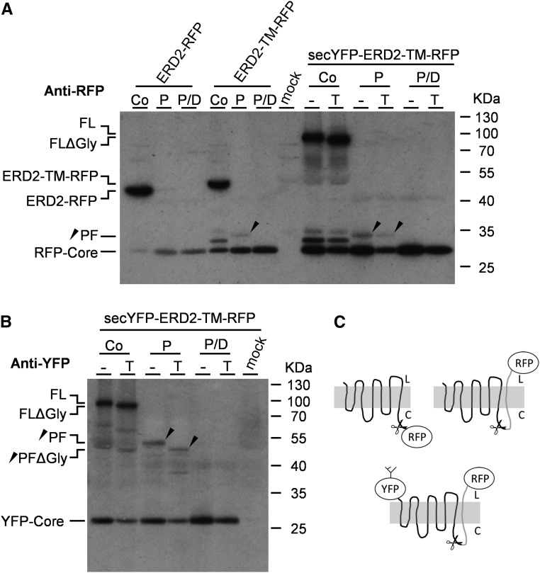
Figure 6.
Experiments Using Modifications of the ERD2 C Terminus.
(A) Protease protection analysis of transiently expressed fusion proteins ERD2-RFP, ERD2-TM-RFP, and secYFP-ERD2-TM-RFP in tobacco protoplasts with (T) or without (−) tunicamycin. Osmotically stabilized cell extracts containing intact microsomes were either untreated (Co) or digested with proteinase K alone (P) or digested together with detergent (P/D). Immunoblots were probed with anti-RFP serum and included a control lane with an extract from mock-transfected cells as negative control (mock). Individual polypeptide bands include the full-length fusion proteins ERD2-TM-RFP and ERD2-RFP, secYFP-ERD2-TM-RFP with (FL) and without glycan (FLΔGly), the specific protease protected fragment (PF), and the RFP core. The positions of the size markers are indicated on the right and given in kilodaltons. The black arrowhead indicates the position of the PF in the relevant lanes.
(B) Protease protection analysis as in (A) but secYFP-ERD2-TM-RFP lanes probed with anti GFP serum. Abbreviations are as in (A).
C) Schematic drawing of the protein fusions ERD2-RFP, ERD2-TM-RFP, and secYFP-ERD2-TM-RFP with their proposed membrane topologies and the site where proteinase K is likely to cleave the fusion protein (scissors). Notice that all further predicted cytosolic loops of ERD2 appear to be resistant to the protease.

