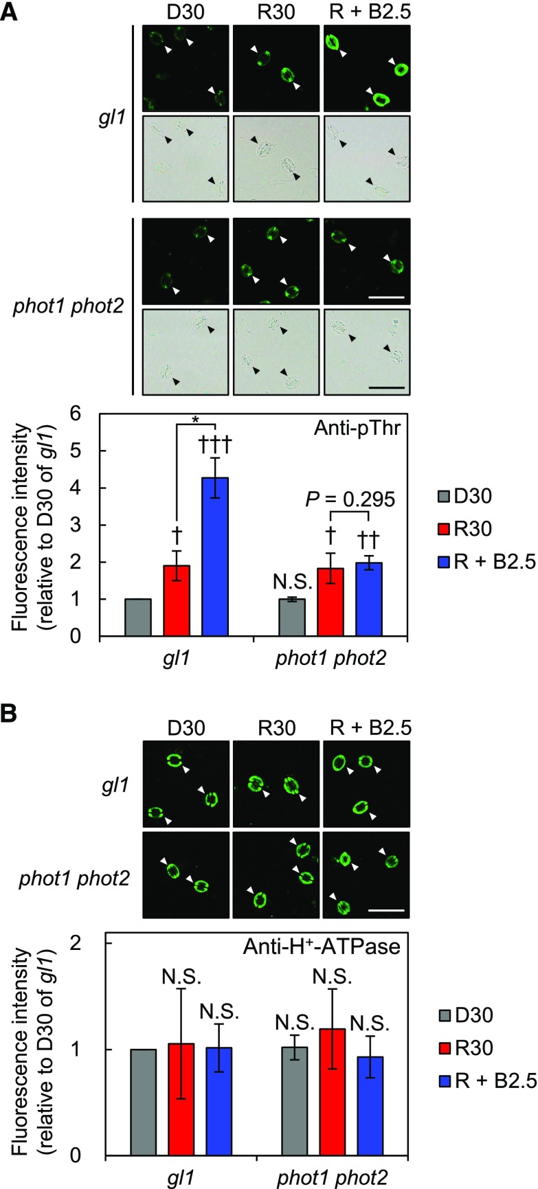
Figure 1.
Establishment of an immunohistochemical technique using whole leaves to detect PM H+-ATPase in guard cells. Phosphorylation (A) and amount of PM H+-ATPase (B) in guard cells in whole leaves from gl1 and phot1 phot2 are shown. Phosphorylation or amount of the protein was detected using anti-pThr or anti-H+-ATPase antiserum, respectively. Mature leaves harvested from dark-adapted plants were illuminated with red light (600 µmol m–2 s–1) for 30 min (R30) followed by blue light (5 µmol m–2 s–1) superimposed on red light for 2.5 min (R + B2.5) or kept in the dark (D30). Typical fluorescence and corresponding bright-field images (top) and quantification of the fluorescence intensities (bottom) are shown. Arrowheads indicate guard cells. Data represent means of relative values from three independent measurements with sd. A, Daggers denote that the mean is statistically significantly higher than D30 of gl1 set to 1. N.S., Not significant (one-tailed one-sample Student’s t test: †, P < 0.05; ††, P < 0.01; †††, P < 0.005; and N.S., P = 0.516). The asterisk indicates that the mean of R + B2.5 is statistically significantly higher than that of R30 in gl1 (one-tailed Student’s test: *, P < 0.005). B, The means were not statistically significantly different from D30 of gl1 set to 1 (two-tailed one-sample Student’s t test: N.S., P > 0.45). Bars = 50 µm.

