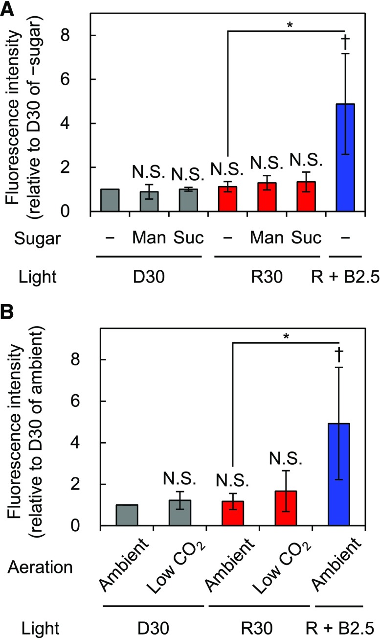
Figure 7.
Effects of Suc or CO2 concentration on the phosphorylation of PM H+-ATPase in guard cells in isolated epidermis. Immunohistochemical detection of the phosphorylation of PM H+-ATPase in stomatal guard cells using anti-pThr antiserum is shown. Epidermal tissue was illuminated with red light (600 µmol m–2 s–1) for 30 min (R30) followed by blue light (5 µmol m–2 s–1) superimposed on red light for 2.5 min (R + B2.5) or kept in the dark (D30) with the indicated conditions. A, Epidermis was incubated without (–) or with 30 mm mannitol (Man) or Suc. B, Experimental basal buffer was aerated with ambient or soda lime-passed low-CO2 air (CO2 = 30–40 µL L−1). The quantification of fluorescence intensities is shown. Data represent means of relative values from independent measurements (A, n = 4; B, n = 5) with sd. Daggers denote that the mean is statistically significantly higher than D30 of –sugar (A) or that of ambient (B) set to 1. N.S., Not significant (one-tailed one-sample Student’s t test: †, P < 0.05; N.S. [A], P > 0.08; and N.S. [B], P > 0.10). Asterisks indicate that the mean of R + B2.5 is statistically significantly higher than that of R30 with a corresponding control condition (one-tailed Student’s t test: *, P < 0.01).

