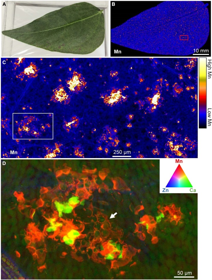
Figure 3.
Mn accumulating in an excised (hydrated) trifoliate leaf of cowpea exposed to 30 µm Mn in nutrient solution. A, Image of the leaf. B and C, Distribution of Mn. D, Tricolor image showing Mn, Ca, and Zn. The area scanned in C is indicated by the red rectangle in B, and the area scanned in D is indicated by the white rectangle in C. In D, note how Mn (red) initially accumulates in the cell wall (white arrow), with the green (Ca) circular structures corresponding to vacuoles. For more details and full experimental procedures, see Blamey et al. (2018a).

