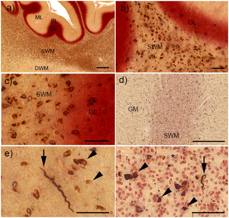
FIGURE 3.
α-Synuclein (α-syn) pathology in the cerebellum and cortex of cases with striatonigral degeneration (SND). (A) Overview of glial cytoplasmic inclusions (GCI) in different layers of the cerebellar cortex. While severe GCI pathology is present in the subcortical white matter (SWM), deep white matter (DWM) and granular layer (GL) are much less involved, and molecular layer (ML) and Purkinje cell layer (PL) show no GCI. (B, C) Higher-resolution images show severe GCI in the SWM and milder pathology in the GL. (D) GCI pathology in the grey matter (GM) and SWM of the orbital frontal cortex. (E) Neuritic inclusions (NRI, arrow) and GCI (arrowheads) in the cortical GM. (F) Neuritic inclusions (NRI, arrow) and GCI (arrowheads) in the SWM, small reddish oligodendroglia without GCI are shown by asterisks. All scale bars = 50 μm; scale bars in (A) and (D) = 500 μm.

