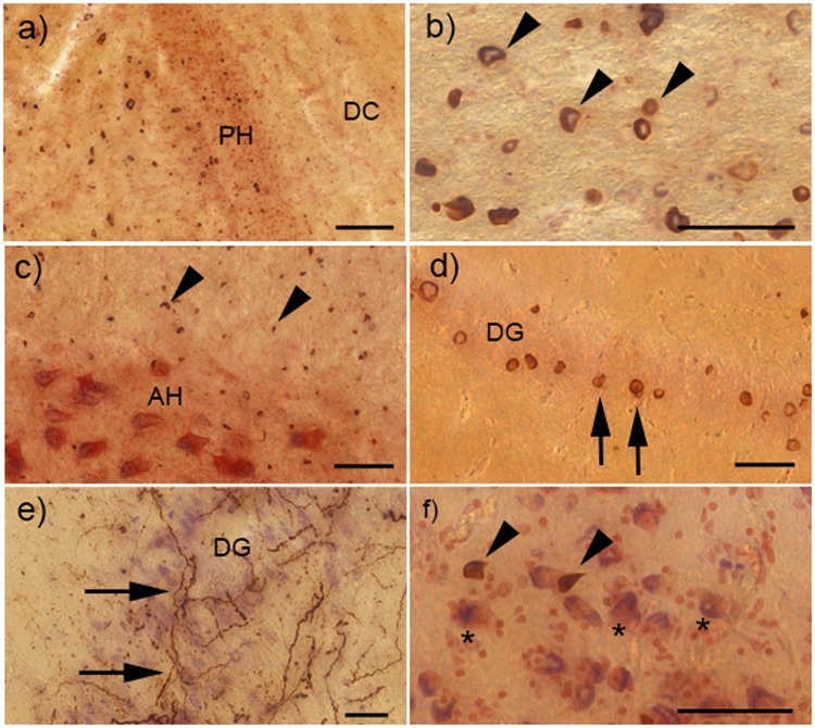
FIGURE 4.
α-Synuclein (α-syn) pathology in the cervical spinal cord, hippocampus, and amygdala of cases with striatonigral degeneration (SND). (A) Shows multiple glial cytoplasmic inclusions (GCI) in the spinal cord white matter, though the dorsal column (DC) to the right of the posterior horn (PH) is only very mildly involved. (B) Multiple GCI (examples depicted by arrowheads) in the corticospinal tract. (C) Multiple GCI in the white matter surrounding the anterior horn (AH), and some GCI within the AH, but no neuronal inclusions (NI) can be seen. (D) NI within the dentate gyrus (DG) of the hippocampus (examples shown by arrows). (E) Severe neuritic inclusions (NRI) within the DG. (F) GCI (shown by arrowheads) among the neurons of the amygdala (examples depicted by asterisks). Scale bar in (A) = 100 μm; all other scale bars = 50 μm.

