Figure 4.
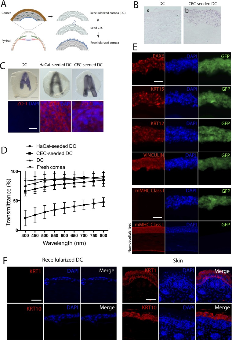
Recellularization of mouse DC with hESC-derived CEC. (A) A scheme for the corneal decellularization and recellularization. (B) H&E staining for determination of the multilayered structure of recellularized DC. Scale bar: 50 μm for both images. (C) Opacity and epithelial cell integrity of the three engineered samples DC, DC recellularized with HaCat keratinocytes, and DC recellularized with CEC. The opacity is reflected as the fuzziness of a letter A underneath the samples (upper), whereas the epithelial cell integrity was detected based on ZO-1 expression in the samples detected via whole-mount immunostaining. Scale bar: 50 μm for all. (D) The average of % transmittance of light at various wavelengths through DC, CEC- and HaCat-seeded DC (n = 3). (E) Fluorescent immunostaining for PAX6, KRT15, KRT12, VINCULIN, and mouse cell-specific MHC class I (mMHC-I) in CEC-seeded DC (upper five rows) with nondecellularized mouse cornea as a positive control for mMHC-I (bottom row). Scale bar: 50 μm for all. (F) Fluorescent immunostaining for keratinocyte markers KRT1 and KRT10 in CEC-seeded DC and sections of mouse skin tissues as a positive control. Scale bar: 50 μm for all.

