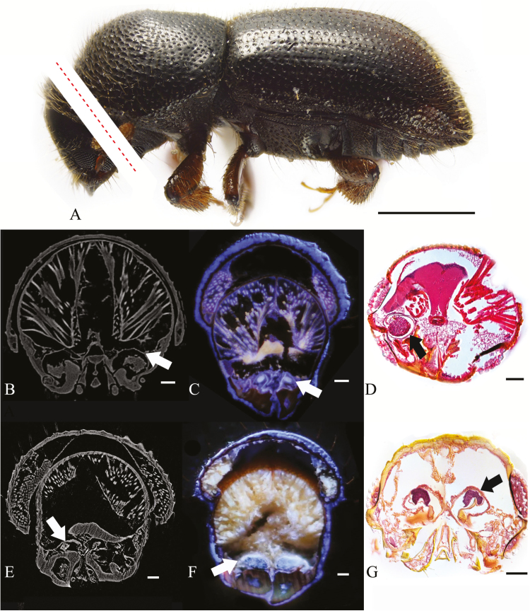
Fig. 1.
Cross-sections of oral mycangia from ambrosia beetle; (A)(C) Ambrosiophiuls atratus; red dotted line digitally illustrating the location of sections only, not actual photographs; (B)(D) Premnobius cavipennis; (E)(G) Ambrosiodmus minor; (F) Euwallacea interjectus; (B)(E) micro-CT; (C)(F) LATscan; (D)(G) paraffin section; white/black arrows: mycangial membrane; scales bar, (A) 1 mm; (B–J) 0.1 mm.

