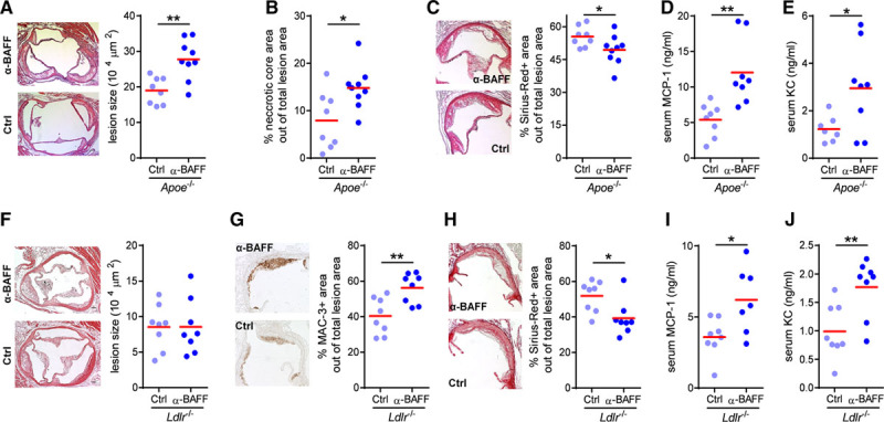
Figure 2.

Anti-B cell–activating factor (BAFF) antibody treatment aggravates atherosclerosis. (A) Representative photomicrographs of hematoxylin and eosin–stained aortic root lesions (50×) and dot plot of the average lesion size in the aortic root expressed as µm2 per section (400 µm), (B) dot plot of the average necrotic area in the aortic root, (C) representative photomicrographs of Sirius Red+ area in aortic root plaques, and serum (D) KC (keratinocyte chemoattractant) cytokine and (E) MCP-1 (monocyte chemoattractant protein-1) chemokine levels of Apoe−/− mice fed an atherogenic diet for 6 weeks and injected with a control (Ctrl) or an anti-BAFF neutralizing antibody (n=8 to 9 mice per group). (F) Representative photomicrographs of hematoxylin and eosin–stained aortic root lesions (50×) and dot plot of the average lesion size in the aortic origin expressed as µm2 per section (400 µm), representative photomicrographs and dot plot of the average (G) MAC-3 and (H) Sirius Red+ area in aortic root plaques, and serum (I) KC cytokine and (J) MCP-1 chemokine levels of Ldlr−/− mice fed an atherogenic diet for 8 weeks and injected with a Ctrl or an anti-BAFF neutralizing antibody (n=7 to 8 mice per group). Dots represent individual mice. All results show mean. *P<0.05, **P<0.01 (Mann-Whitney U or unpaired Student’s t test). Scale bar: 200 µm.
