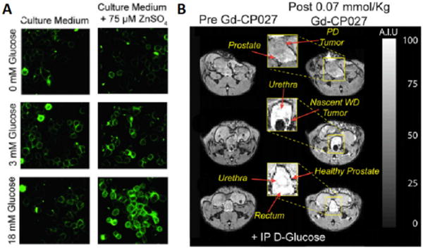
Figure 7.

(A) In vitro fluorescence images of prostate epithelial cells (RWPE-1) cultured in a low-glucose medium without (Left) or with (Right) 75 μM ZnSO4. 1 μM ZIMIR was added to label the outer surface of the cells for detection of zinc release from intracellular stores. Only cells that had accumulated substantial zinc during incubation released zinc in response to glucose in a concentration-dependent manner. (B) Glucose-stimulated contrast enhanced T1-weighted 3D MR images at 9.4 T of the TRAMP mouse prostate during various stages of tumor development. (Bottom) A typical glucose stimulated zinc secretion pattern of the healthy prostate in a young TRAMP mouse (age: 10 wk). (Middle) A nascent tumor in dorsal lobe of the prostate is hypointense presumably reflecting a reduction in the release of intracellular zinc (age: 17 wk). (Top) A poorly differentiated (PD) tumor that originated in lateral lobe and extending to ventral lobe. No glucose stimulated zinc secretion was detected in the tumor, and significant loss of noncancerous prostate was observed (age: 19 wk). GdDO2A-diBPEN-propionate as shown in Figure 6 was used in this study. Reproduced with permission from Proceedings of the National Academies of Science.[38a]
