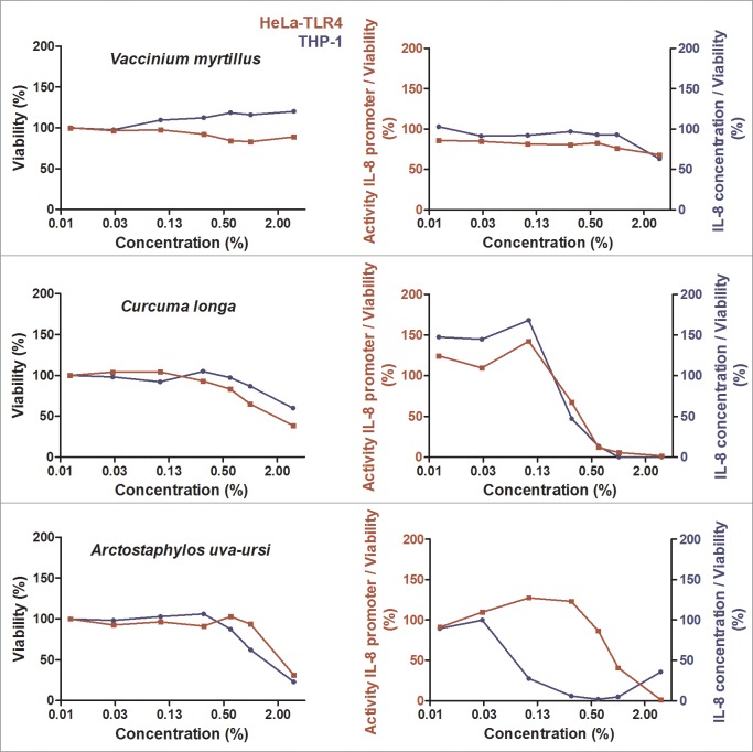
Fig 3. Cell viability and concentration-dependent anti-inflammatory effects of selected herbal extracts (part 3).
HeLa-TLR4 cells (red) and THP-1 monocytes (blue) were incubated with extracts in different concentrations or vehicle (70% ethanol), followed by stimulation with LPS-EB. Viability was measured using the Alamar Blue Assay and was normalized to the negative control (untreated cells). TLR4 receptor stimulation was measured using Renilla luciferase expression for the HeLa-TLR4 cell line and IL-8 ELISA (pg/ml) for the THP-1 monocytes and was normalized to ethanol-treated cells. Data are displayed as viability (%) in the left graphs and TLR4 stimulation divided by normalized viability (%) in the right graphs. Data represents means (n≥2). For graphical display of further extracts, see Fig 1, Fig 2 and supplementary data S1 Fig.

