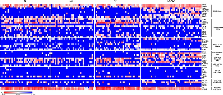
Fig 4. Three TF combinations induce hES-iNs with similar molecular signature resembling that of fetal human cortical neurons.
Single-cell quantitative RT-PCR analysis of expression levels of different cortical genes listed to the right. Expression levels are shown as Ct values and color-coded as shown on the bottom. The mRNA levels were quantified in hES-iN cells, co-cultured with primary mouse astrocytes for 8 weeks and FACS-sorted as NCAM+, and in hFCtxF cells from a 7 weeks old human fetus.

