Figure 3.
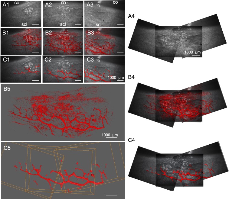
Rendering and alignment of adjacent outflow tract vessel volumes in 3D. Volume intensity projections (VIPs) (A1–3) of adjacent OCT scans with minimally processed (B1–3) and cleaned signal voids (C1–3) in overlay. (A4–C4) 2D alignment of images in (A–C), 1 to 3 fails to account for the curvature of the ocular surface, resulting in alignment artifacts. (B5) surfaces in (B1–3), and (C5) in (C1–3) are aligned in 3D space to neighboring scans without artifacts. Bounding box (orange) coordinates in (C5) were conferred to respective scans in (B5) to align them. co, cornea; scl, sclera.

