Figure 6.
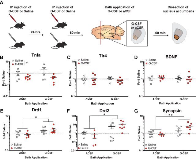
G-CSF treatment reduces Tnfa expression and primes postsynaptic signaling. A, Timeline of G-CSF injections. Animals were injected with either saline or G-CSF 24 h and then 60 min (left) before G-CSF or aCSF bath application and dissection of NAc (right; n = 5–7 per group). B, NAc Tnfa mRNA levels calculated as fold-changes from the Saline/aCSF-treated controls. Tnfa expression in the NAc was decreased in the G-CSF pretreated group regardless of bath application. C,Tlr4 expression in the NAc was not affected by G-CSF pretreatment or bath application. D, Bdnf expression in the NAc was not altered by G-CSF. E, NAc Drd1 expression levels represented as fold-changes from saline/aCSF-treated controls. F, NAc Drd2 mRNA levels. G, Syn3 expression in the NAc. Expression levels of all three genes were elevated following G-CSF bath application regardless of pretreatment.*p < 0.05, **p < 0.01 between bath applications. All data are presented as mean SEM.

