Figure 5.
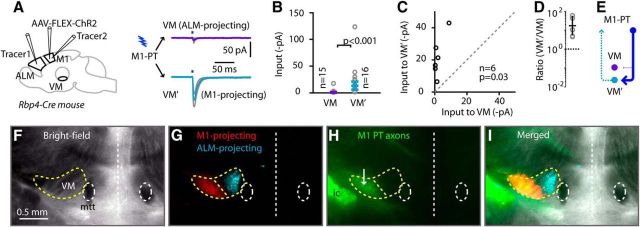
M1 PT axons excite M1-projecting VM TC neurons, but not ALM-projecting VM TC neurons. A, Left, Schematic summary of injection strategy. Right, Average responses (±SEM; gray lines) recorded in ALM-projecting and M1-projecting VM neurons. B, Cell-based group comparison. C, Animal-based group comparison. D, Animal-based ratios. E, Schematic depiction of the cellular connectivity pattern. F, Bright-field view of thalamic slice, showing VM located dorsolateral to the mammillothalamic tract (mtt). G, Merged fluorescent image showing thalamic distributions of ALM-projecting (cyan) and M1-projecting (red) neurons in medial and lateral, respectively, parts of VM. H, Fluorescence image of ALM PT axons (green) following injection of AAV-FLEX-ChR2-tdTomato into M1 of an Rbp4 animal. ic, Internal capsule. I, Merged image of all three channels.

