Figure 3.
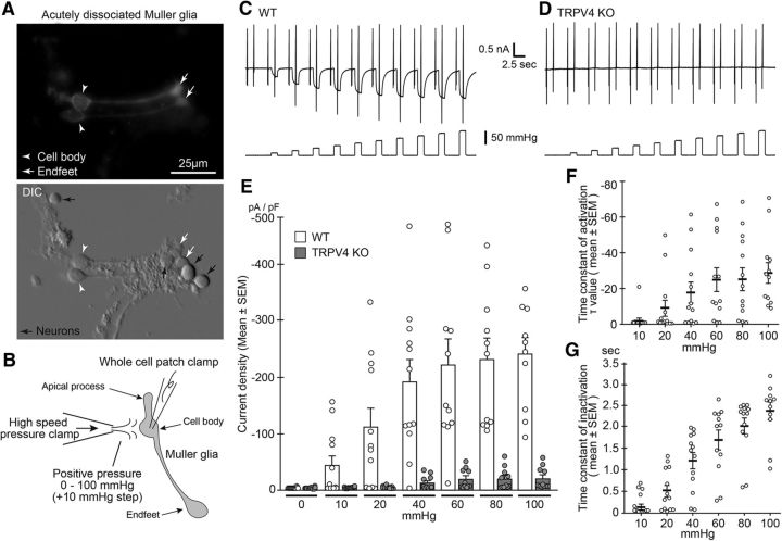
Müller glial TRPV4 is activated by mechanical stimuli. A, Morphology of acutely dissociated Müller glial cells. White arrowheads and arrows represent soma and endfeet, respectively. Top, Immunostaining of acutely dissociated cells with glutamine synthetase (a Müller glial marker). Black arrows represent neurons. B, Schematic drawing of a Müller glial cell and electrophysiological recording by stepped positive pressure stimulation (+10 mmHg per step). C, D, Representative traces of WT or TRPV4KO cells after application of stepped positive pressure with ramp pulses (−100 to +100 mV). The holding potential was at −60 mV. E, Quantification of densities of positive pressure-evoked currents. WT, n = 10; TRPV4KO, n = 12. F, Quantification of time constant of activation periods in WT recording (n = 10). G, Quantification of time constant of inactivation periods in WT recording (n = 10).

