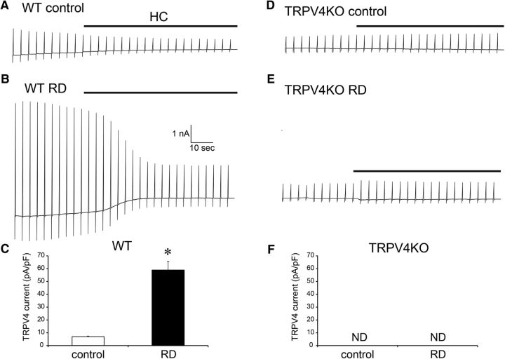
Figure 5.
RD-induced Müller glial swelling activates TRPV4 at body temperature. A, B, Representative traces of Müller glia in a WT retinal explant at 37°C. The recording pipette was placed in the endfeet. The holding potential was at −60 mV. Ramp pulses (−100 to +100 mV) were applied in each 5 s interval. The black lines represent application of a TRPV4 antagonist HC (10 mm). Control is from normal retinal explants. RD is from RD-evoked retinal explants. C, Quantification of densities of TRPV4-evoked currents in WT explants (n = 5, *p = 0.0058, Student t test). D, E, Representative traces of Müller glia in TRPV4KO a retinal explant at 37°C. The recording pipette was placed in the endfeet. The holding potential was at −60 mV. Ramp pulses (−100 to +100 mV) were applied in each 5 s interval. The black lines represent application of a TRPV4 antagonist HC (10 mm). Control is from normal retinal explants. RD is from RD-evoked retinal explants. F, Quantification of densities of TRPV4-evoked currents in TRPV4KO explants (n = 5). We failed to observe any TRPV4 currents.

