Figure 8.
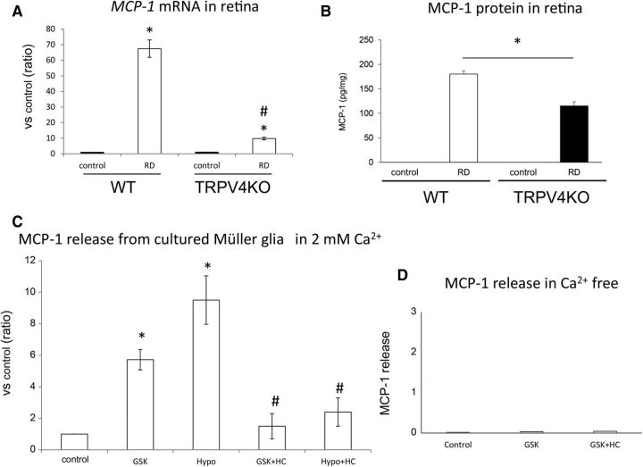
TRPV4 activation triggers MCP-1 release from Müller glial cells and leads to photoreceptor apoptosis in the detached retina. A, Real-time PCR assays were performed using specific MCP-1 primers. The values were normalized against β-actin expression, and the bar graphs compare values with the MCP-1/β-actin expression level of control eyes in WT mice. Asterisks (vs control) and hash tags (vs WT RD) represent a significant difference at p < 0.01 (*p = 0.000116, #p = 0.000232, Student t test, n = 5). B, ELISAs of MCP-1 protein were performed in control and RD eyes for WT and TRPV4KO mice. The asterisk represents a significant difference at p < 0.05 (p = 0.0010509, Mann–Whitney U test, n = 6). C, ELISA assays of MCP-1 protein performed using cultured Müller glial cells from WT mice. Asterisks (vs control) and hash tags (vs GSK or Hypo) represent a significant difference at p < 0.01 (*GSK, p = 0.000247; *Hypo, p = 0.00944; #GSK+HC, p = 0.001683; #Hypo+HC, p = 0.00092; t test, n = 4). D, ELISAs of MCP-1 protein were performed using the supernatant of cultured Müller glial cells (from WT mice) under Ca2+-free conditions. Only the small amount of MCP-1 release was observed independent from TRPV4 activation or inhibition (GSK or HC). These results were perfectly different from the physiological Ca2+ condition (2 mm) shown in Figure 5C. Thus, we can conclude that TRPV4-triggered Ca2+ influx evokes the MCP-1 release from Müller glial cells.

