
Figure 2.
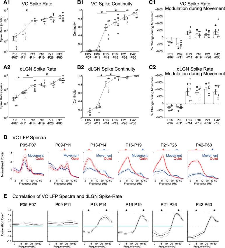
Continuous activity, spike rate, and modulation during movement develop simultaneously in thalamus and cortex. A, Spike rate of VC (A1) and dLGN (A2) increases throughout development. Circles show multiunit cortical layer 4 (A1) and dLGN (A2) spike rates from each animal taken from all conditions (quiet and moving). Line and error bars depict mean ± SEM from n = 6 animals in each age group. Asterisk line shows result of post hoc test following one-way ANOVA for age (p < 0.05). Only the shortest significance is shown for clarity (e.g., P9–P11 is also different from P16, P21, and P42 groups). B, Continuity of MUA in L4 VC (B1) and dLGN (B2) increases throughout development, largely between P11 and P13. C, MUA L4 VC and dLGN firing-rates switch from negatively to positively correlated with movement between P11 and P13. Percentage change of VC (C1) and dLGN (C2) spike rate during movement relative to quiescence is shown at each age (asterisk marked arrows show significant difference from zero (p = 0.031) by one-sample Wilcoxon signed rank test. D, Adult-like modulation of the VC LFP during movement emerges at P13. Population mean (n = 6 each) VC LFP spectra during movement (blue, solid line) and quiescence (red, dashed line) are shown for each age group. Thin lines show SEM. Frequency ranges with significant increase during movement are marked by blue bars; during quiescence by red bars (permutation test, *p < 0.05). E, Adult-like correlation between VC LFP power and dLGN firing-rate emerge after P13. Frequency ranges significantly different from zero are marked by black bars (permutation test, *p < 0.01).