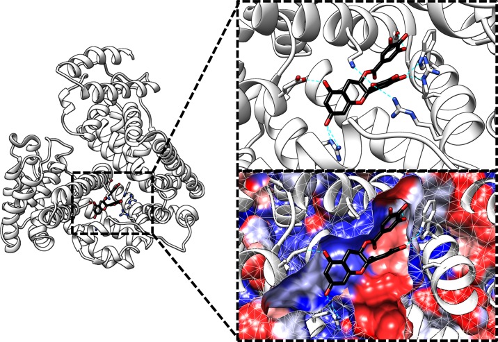
Fig 4. Model of a complex between human serum albumin (white) and (−)-epigallocatechin gallate (black) obtained by means of a docking simulation.
Only residues that formed hydrogen bonds with EGCg were shown. Hydrogen bonds were described as cyan dotted lines. The molecular surface of the protein was colored based on the Kyte–Doolittle hydrophobicity scale (blue for hydrophilic areas and red for hydrophobic areas [38]). The figure was made by the UCSF Chimera [46].

