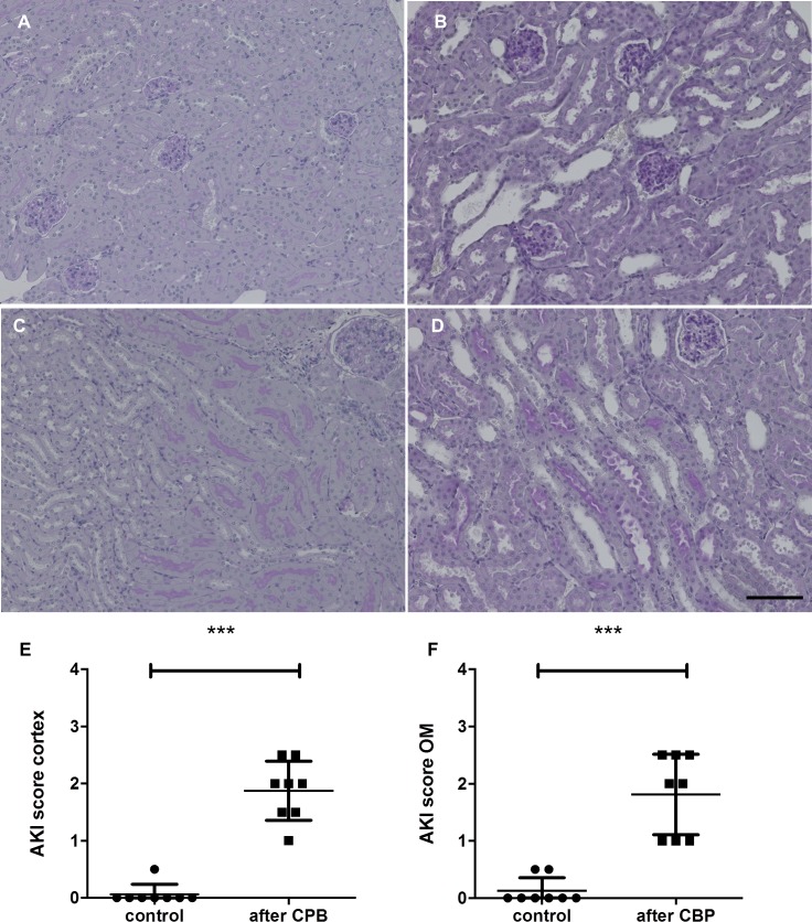
Fig 1.
PAS staining of the kidney after CPB with MHCA in (A, C) healthy control animals and (B, D) after CPB. The upper row shows the cortex, the lower row the outer medulla. Kidney morphology after CPB showed acute tubular damage with tubular dilation. Semi-quantitative analysis was done for the cortex and outer medulla separately (E, F, ***P<0.001). Scale bars, 100 μm.

