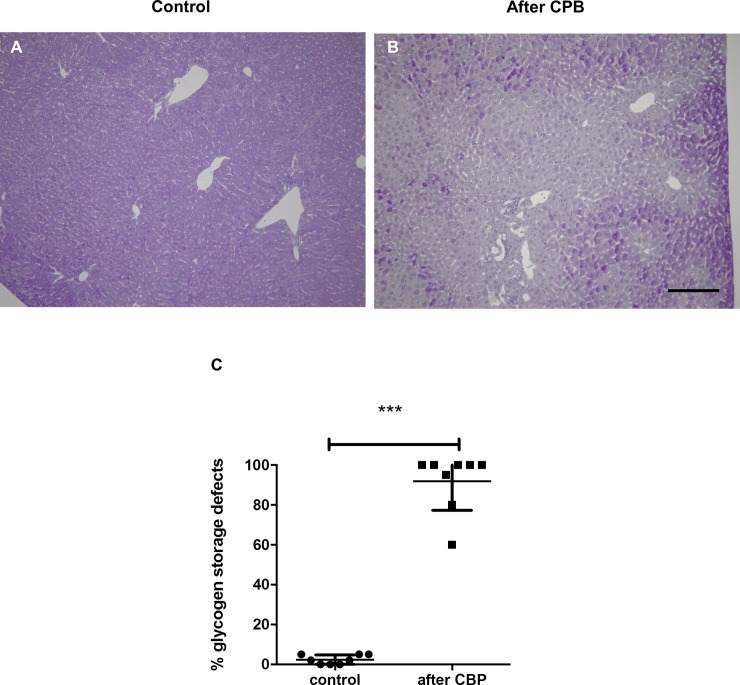
Fig 3. PAS staining of the liver reveals loss of glycogen storage capacity after CPB with MHCA.
(A) Representative liver sections of healthy controls and (B) CBP/MHCA experimental mice. Dark purple staining indicates normal glycogen storage capacity and pale areas indicate loss of glycogen storage in injured hepatocytes. The proportion of hepatocytes demonstrating loss of glycogen storage capacity was quantified. CBP caused reduction of glycogen storage capacity (C, ***P<0.001). Scale bar, 100 μm.

