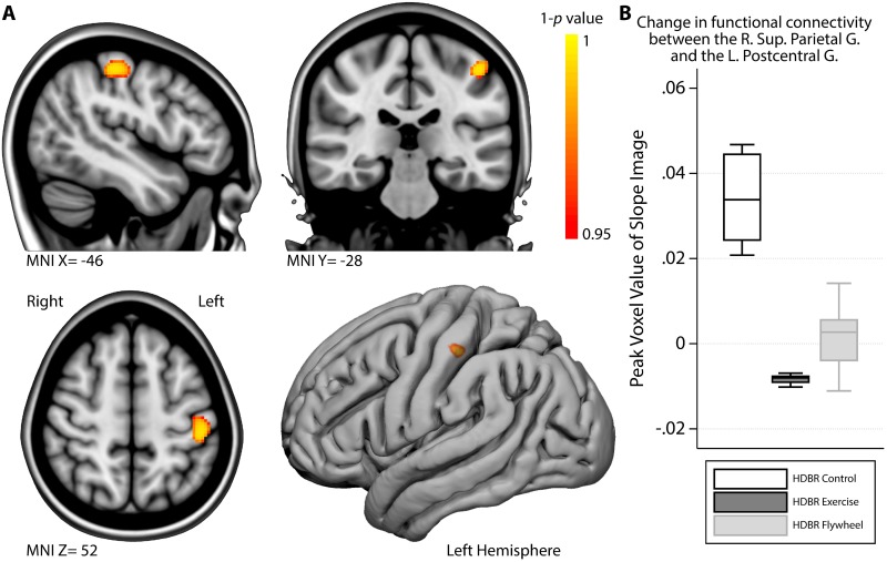
Fig 3. Exercise modulates functional connectivity strength between the right superior parietal gyrus and the left postcentral gyrus post-HDBR.
A) Regions in which HDBR control subjects showed a significantly larger increase in functional connectivity between the Right Superior Parietal Gyrus and the Left Postcentral Gyrus than the HDBR exercise subjects during the post-HDBR phase. B) Boxplot of the functional connectivity strength measure (daily change in correlation) of the most significant ‘peak’ voxel in the area depicted in A, stratified by group. MNI = Montreal Neurological Institute coordinate; R. = Right; L. = Left; G. = Gyrus.

