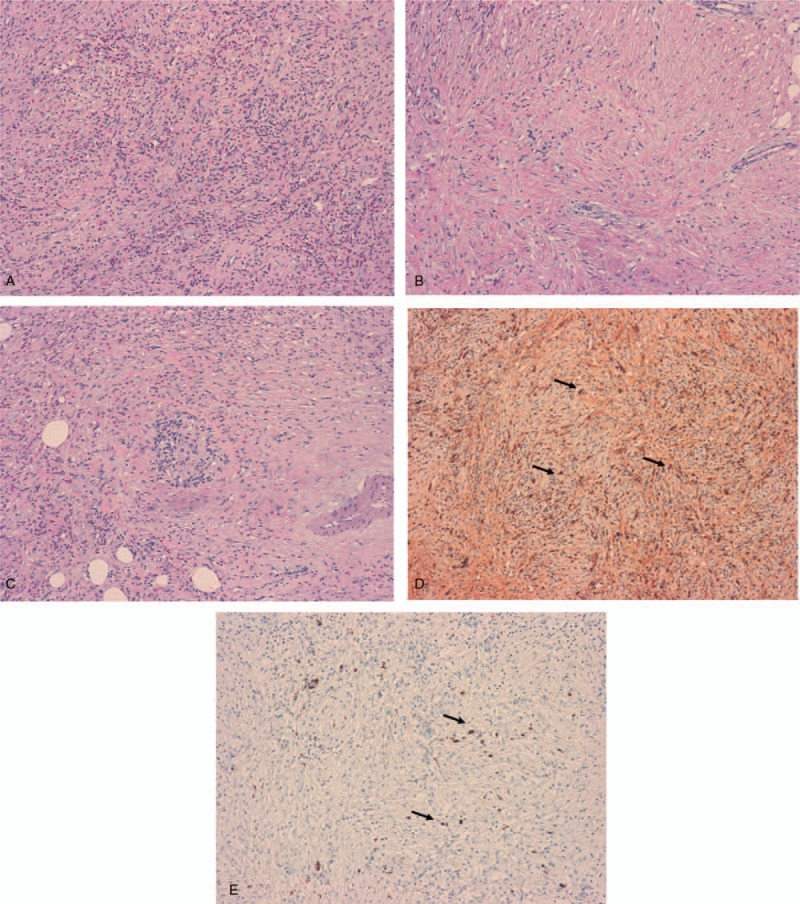
Figure 4.

Histological and immunochemical analysis of the biopsies from the abdominal mass. (A) Severe inflammatory infiltration of the mesentery made of eosinophils, lymphocytes and plasma cells (hematoxylin eosin staining, H&E, ×10 magnification). (B) Histological section (H&E, × 10 magnification) showing a storiform pattern of fibrosis. (C) The vein in the center of the image is obliterated by the infiltration of inflammatory cells (obliterative phlebitis). (D) IgG-immunostaining (×10 magnification) showing IgG positive plasma cells (arrows). (E) IgG4-immunostaining (×10 magnification) showing IgG4 positive plasma cells (arrows).
