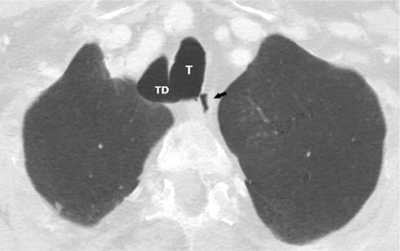
Figure 2.

Contrast-enhanced MDCT, 1.5 mm thickness, lung window, MinIP. Axial images of the chest showing a large TD located at the right paratracheal area (T). Black arrow shows esophagus. MDCT = multidetector computed tomography, MinIP = minimum intensity projection, TD = tracheal diverticulum, T = trachea.
