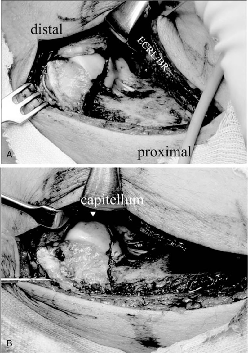
Figure 2.

A, An anterolateral approach clearly exposes the anterior aspect of the articular surface of the lateral condyle. B, Following anatomical reduction, 2 Kirschner wires are inserted from the lateral condyle under fluoroscopic guidance. BR = brachioradialis, ECRL = extensor carpi radialis longus.
