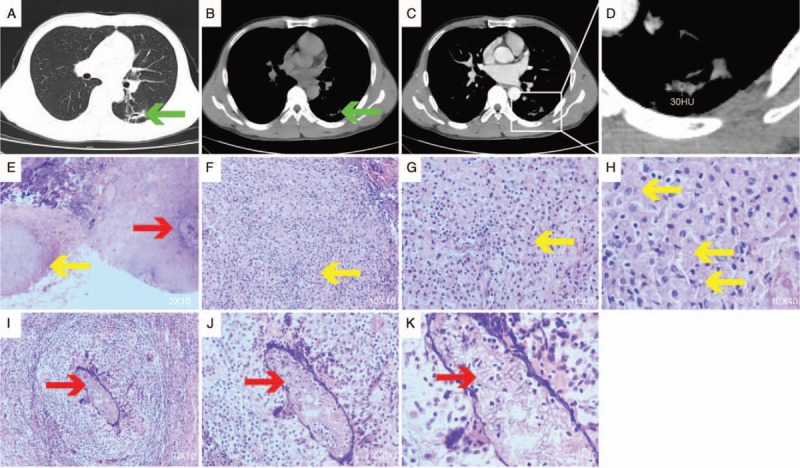
Figure 1.

Chest radiological changes and pathological findings. (A) 3.0-cm increase in density with clear boundaries in the left upper lobe; (B, C) enhanced and non-enhanced computed tomography (CT) images of 2 nodes of the pulmonary mediastinal window; (D) enlarged view of the pulmonary mediastinum window nodules showing 2 nodules with irregular and well-defined borders in the upper left lobe. The larger nodule has an irregular border with a uniform density of 2.4 cm, in which cavities are visible. The smaller 0.7-cm nodule is on the left; (E) surgically resected lung biopsy. Pathology suggested that both fungal foci existed separately (hematoxylin and eosin stain, original magnification 20×). The yellow arrow tips are cryptococcal lesions, and the red arrow tips are aspergillosis lesions; (F–H) visible cryptococcosis; (I–K) Aspergillus can be seen, and there is a large number of chronic inflammatory cells and histiocytic infiltration, lymphoid follicle formation, and a large amount of local necrosis (hematoxylin and eosin stain, original magnification 100×, 200×, 400×).
