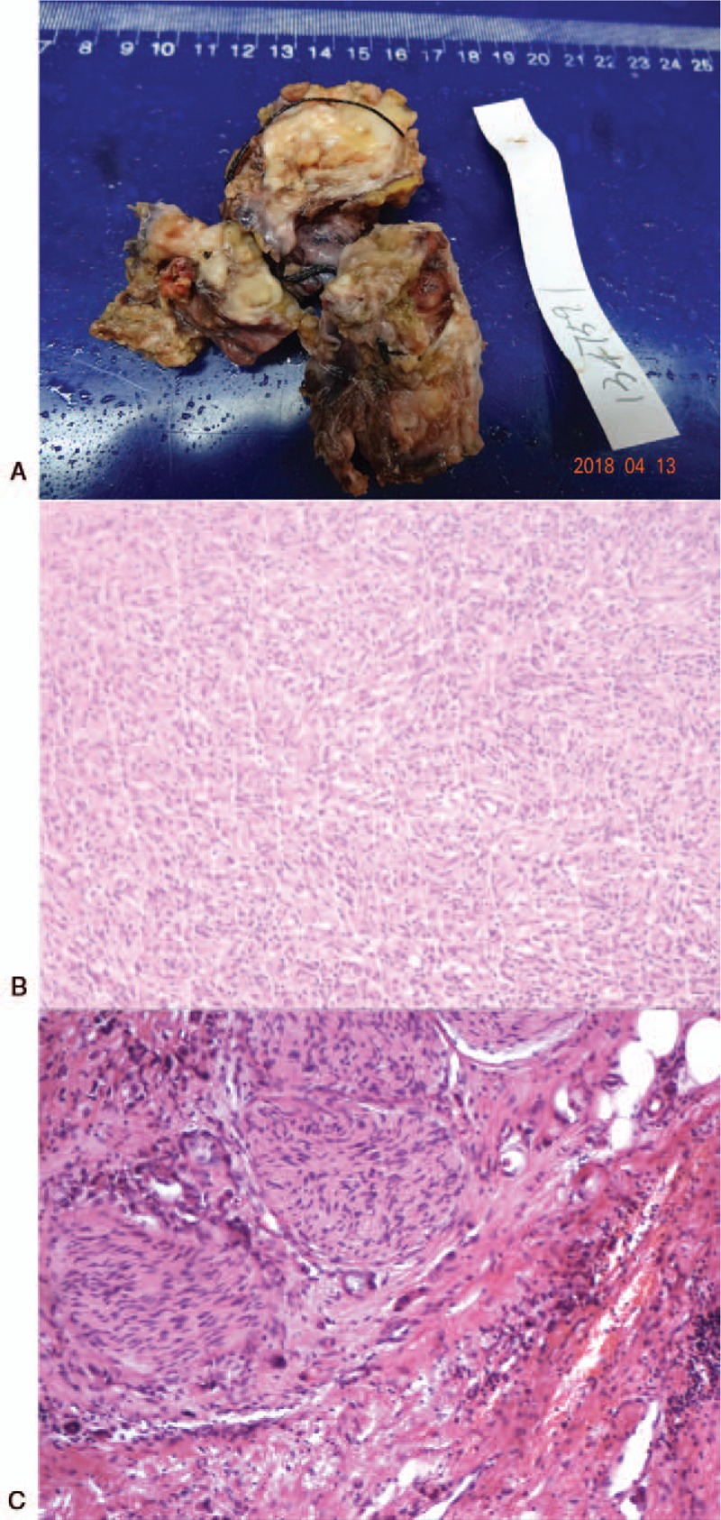
Figure 2.

Morphological and histological appearance of the tumor, which consists of Sarcomatous components, with heterogeneous differentiation (chondrosarcoma). (A) The surgical specimens of the liver mass. (B) HE staining of the tumor on left lateral lobe of the liver (60×). (C) HE staining of the tumor on left lateral lobe of the liver (150×). HE = hematoxylin and eosin.
