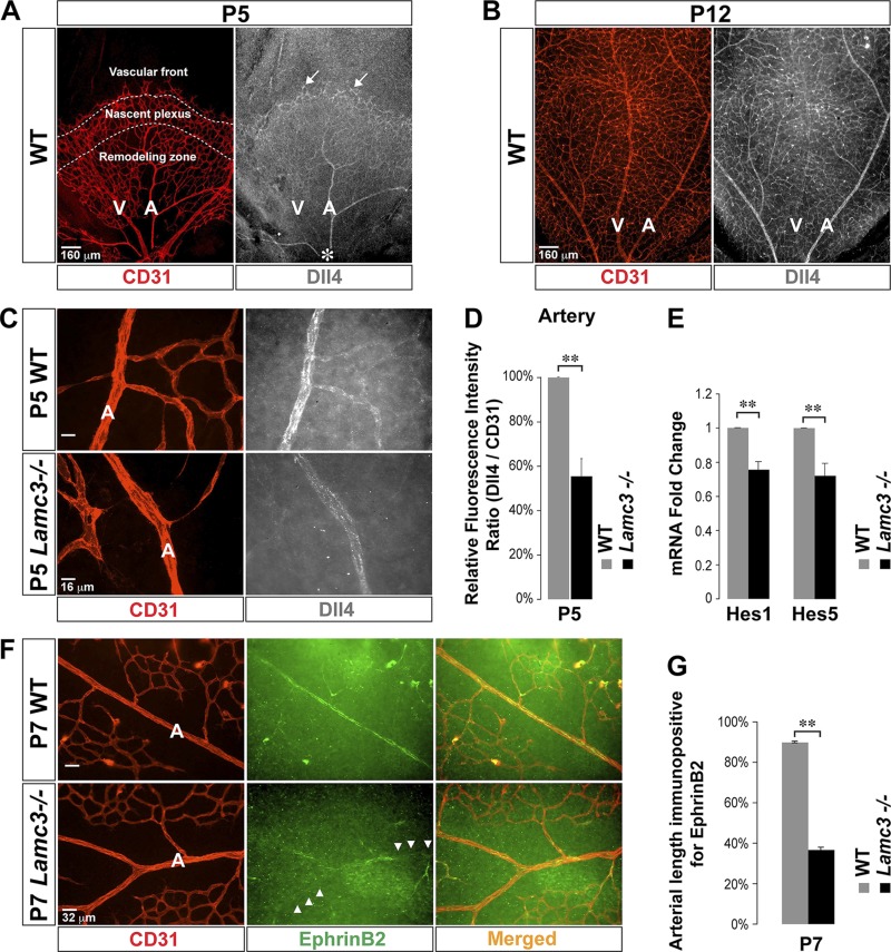
Figure 1.
Arterial Dll4/Notch signaling is disrupted in the Lamc3−/− retina. A) CD31 (EC marker: red) and Dll4 (white) labeling demonstrated high Dll4 expression in the tip cells (arrows) at the vascular front (P5). There was little Dll4 expression in the nascent plexus and the Dll4 expression pattern became artery specific in the remodeling zone as arteries mature from behind the nascent plexus toward the optic nerve head (asterisk). B) The artery-specific expression pattern of Dll4 persists in the mature superficial vasculature (P12). C) Higher power images of CD31 (red) and Dll4 (white) labeling of arteries; arterial Dll4 expression was down regulated in the Lamc3−/− retina. D) Dll4 immunofluorescence was quantified relative to CD31 fluorescence, demonstrating a significant decline in P5 Lamc3−/− arteries (n = 3). E) mRNA expression of Notch target genes; Hes1 and Hes5 mRNA levels were down-regulated in the Lamc3−/− retina (n = 3). F) CD31 (red) and Ephrin-B2 (green) labeling of arteries demonstrated that Lamc3−/− arterial regions completely lacked Ephrin-B2 expression (arrowheads). G) Quantification of arterial length immunopositive for Ephrin-B2 demonstrated significantly less extensive arterial Ephrin-B2 immunoreactivity in the Lamc3−/− retina (n = 2). A, artery; NS, not significant; V, vein. Scale bars: 160 μm (A, B), 16 μm (C), 32 μm (F). Data are means ± sem. **P < 0.02.

