Figure 2.
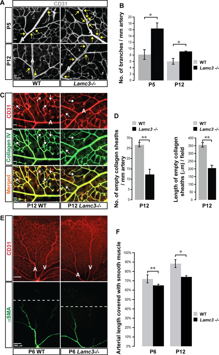
Arterial morphogenesis is disrupted in the Lamc3−/− retina. A) CD31 (white) labeling demonstrates more primary arterial branches (arrows) in the Lamc3−/− retina. B) Arterial branching is quantified demonstrating a significant increase in the Lamc3−/− retinae (n = 3). C) CD31 (red) and collagen IV (green) labeling of arteries demonstrates less extensive vascular pruning around Lamc3−/− arteries. Arrows: empty collagen sheaths (collagen IV+/CD31−) demarking pruned vessels. D) Number and lengths of pruned vessels are quantified demonstrating a significant decrease around Lamc3−/− arteries (n = 3). E) CD31 (red) and α−SMA (green) marker labeling demonstrates less extensive vascular smooth muscle coverage of Lamc3−/− arteries. Dashed lines: extent of arterial smooth muscle coverage. F) Quantification of the extent of arterial smooth muscle coverage, relative to vascular migration distance, demonstrates significant reduction in Lamc3−/− arteries (n = 3). A, artery; V, vein. Scale bars: 32 μm (A, C), 160 μm (E). Data are means ± sem. *P < 0.05, **P < 0.02.

