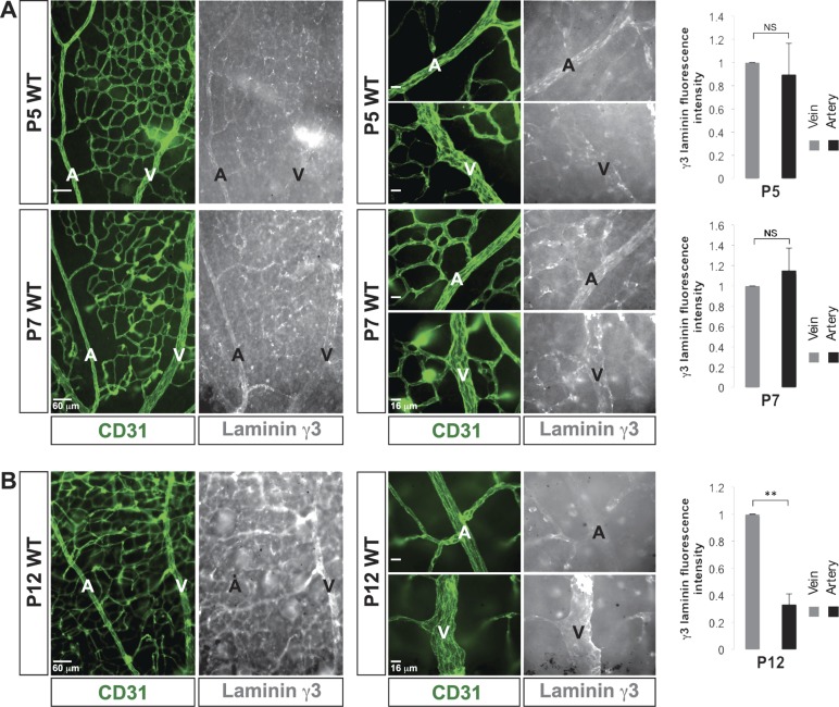
Figure 3.
γ3-Laminin is deposited in both arterial and venous BMs during retinal arteriogenesis. A) CD31 (green) and γ3-laminin (white) labeling demonstrates that γ3-laminin was deposited in emerging arterial, venous, and microvascular BMs during retinal arteriogenesis (P5 and 7). Quantifications showed no difference in relative expression of γ3-laminin between arteries and veins at these time points (n = 3). B) In mature vessels (P12), γ3-laminins were present, mostly in venous and microvascular BMs. Quantifications showed a drastic decrease in relative expression of γ3-laminin in arteries at that time (n = 3). A, artery; NS, not significant; V, vein. Scale bars: 60 μm, 2 left columns; 16 μm, 2 right columns (A, B). Data are means ± sd. **P < 0.02.

