Figure 6.
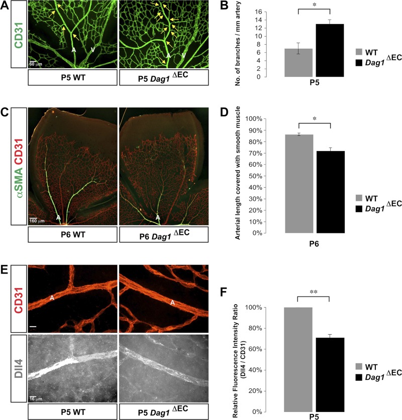
EC-specific deletion of DG gene reproduces Lamc3−/− retinal arterial phenotype. A) CD31 (green) labeling demonstrated more arterial branches (arrows) in the Dag1ΔEC retina at P5. B) Quantification of arterial branching demonstrated a significant increase in Dag1ΔEC retinae (n = 3). C) CD31 (red) and αSMA (green) labeling demonstrated less extensive vascular smooth muscle coverage of Dag1ΔEC arteries at P6. D) Quantification of the extent of arterial smooth muscle coverage, relative to vascular migration distance, demonstrated significant reduction in Dag1ΔEC arteries compared to the littermate WT control (n = 3). E) CD31 (red) and Dll4 (white) labeling of arteries demonstrated down-regulation of arterial Dll4 expression in the Lamc3−/− retina. F) Quantification of Dll4 immunofluorescence relative to CD31 fluorescence demonstrated a significant decline in P5 Dag1ΔEC arteries (n = 3). A, artery; V, vein. Scale bars: 60 μm (A), 160 μm (C), 16 μm (E). Data are means ± sem. *P < 0.05, **P < 0.02.

